Today, panoramic X-rays continue to offer the odontologist an excellent solution for getting to know all the details of the patient's mouth. Despite the great opportunities they offer, it is also possible that a number of problems may arise during the x-ray and its full potential may not be exploited. There are a few basic steps to a good panoramic X-ray that are applied to all X-rays. In addition, there are a number of very common problems, so we will tell you what they are and how best to solve them.
Step 1: Load the cartridge.

An extraoral film, consisting of two fluorescent screens, is used for panoramic X-rays. Each fluorescent screen, when struck by X-rays, forms an image on the film. These screens are 10 to 60 times more sensitive to x-rays than films, so a very low dose of radiation is needed.
Most common cartridge loading problems:

- A small black spot on the corner of the x-ray.
One of the possible causes is that the cassette is damaged or that the film has been exposed to light. The fastest way to correct it is to replace the damaged cassette. We recommend that cassettes be checked frequently for light pressure.
- Part of the image is not visible on the x-ray.
La causa de este error es que la radiografía está invertida, por lo que el profesional dental debería dar la vuelta y colocarla correctamente.
- White spots on the image.
If you ever get a whitish X-ray or a white spot on the image, it is because the screen is scratched. It is important that you handle the screens carefully and use the appropriate cleaning materials for this material to avoid scratches.
- Dark marks or excessive brightness.
This is caused by static electricity. You should remove the film from the cassette. In order to solve this error it is advisable to use humidifiers.
- Multiple images.
Multiple images are produced when there is a double exposure, so it is important that after each exposure, you remove the film.
Step 2: Adjustments to the exposure factors.
Many of the newer rayos X panorámicos automatically set exposure factors by reading a small portion of the X-rays at the start of exposure. However, with most panoramic machines, exposure must be established based on the patient's age and height. Normally, small, medium, or large icons are used.
Since the density of the patient's bone does not always match his physical height, the best help is to see the patient's wrists and ankles. Thick wrists can imply a heavy bone density; age is another factor to take into account, and if the patient is toothless and overweight.
Most common problems with exposure factor settings:
- Light, a pale film with a few black areas
One of the most frequent causes is low exposure. To correct this, you must increase Ma or Kv or use a larger configuration on the machine. We advise you to exclude worn or inverted screens.
- Dark films with loss of detail, amalgams and over-exposure of areas that are still transparent.
The main cause of this error is that the films have too much exposure. To correct this, it is important that you reduce the configuration of the machine. We advise you not to confuse this error with fogged films. Fogged films are distinguished because they have a total grey colour.
Step 3: Remove the patient's jewelry and put on protective aprons
Prior to exposure, the patient should remove all jewelry from the head area. Because earrings, necklaces or other jewelry such as piercings on the tongue or nose will be visible on the x-ray.
More common problems:

- Formation of "ghost" images
These images are produced when an object is captured twice (repeatedly), once by the normal size in the area of rotation of the beam, and others by the opposite area. Ghost" images are easily identified by being on the opposite side of the real image, at the top of the film, and are horizontally striped. They can be easily confused by pathology when reaching the chest area. If a metal apron is used during the exposure process, it must be positioned correctly.
Protective aprons should be used to cover the patient's back and shoulder area. The apron should not extend over the neck or will appear on the film as an opaque "shark fin" object.
Step 4: Bite the bar.
In panoramic X-ray machines, a plastic bite bar with small slots is used to position the anterior part of the patient's teeth in the focal channel. Most machines also offer a guide that is placed against the patient's chin or under the nose. These guides are very useful and not using them can cause anterior-posterior errors.
Most common bar bites problems.
- Blurred front teeth, too small and too narrow.
This problem is caused because the patient's bite is too far from the bite bar. To solve it, you must make sure that the anterior teeth are located in the grooves.
- Blurred and wide anterior teeth, "ghosts" in the jaw and spine.
The cause of this problem is that the patient is biting too far from the bar or is not biting at all.
Step 5: Adjust the chin.
In panoramic x-rays patients should look slightly down, at a point on the floor about 8 feet in front of them, which causes the patient to elevate the palate.
Most common problems with chin adjustment:
- Blurred lower incisor roots. V-shaped jaws.
This problem is caused by the patient's chin being too far down. We recommend its replacement, using correctly the guides for the machine.
- Blurred maxillary incisors, hard palate superimposed on the roots or wide and flat jaw.
This time, the problem is caused because the patient's chin is too far up. As in the previous case, it is advisable to reposition the patient.
Step 6: Positioning in the guides.
All panoramic X-rays have guides or positioning lights to align the patient's digital median plane. It is important that the patient faces forward, without inclination..
Most common errors in the positioning of the guides:
- The teeth are enlarged on one side and narrowed on the other or the nasal structures are not clear.
The main cause is that the patient's head is twisted in the machine. Causing asymmetry. Dentists should use correct guides for that machine.
Step 7: Keep the patient straight.
The patient should be straight to prevent bowing of the neck. The best method of achieving this is to keep the patient away from the bite bar. Have the patient step forward after biting the bar.
Most common problem:
- A white line in the middle of the image
Caused by a "ghost" of the spine caused by the fall. To fix this, ask the patient to step forward and stretch the neck.
Step 8: Get a shade by placing the tongue in the roof of the palate.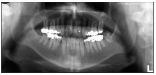
Just before exposure, you should ask the patient to place the tongue in the sky of the mouth and keep it that way during exposure. A small movement or an incorrect position can cause a shadow in the place of the tongue.
Step 9: Developing the film.
Problems during developing are due to machine damage or operator errors. Among these errors, the most common are: releasing the exposure button temporarily (not possible with newer machines), changing the exposure setting during exposure, or not having the cassette properly inserted into the machine. Cassettes should be inserted with the smooth, flat side facing the X-ray tube.

In addition, it is recommended that you check your X-ray machines frequently. There are many options on the market, such as panoramic X-rays, which thanks to this panoramic radiography technology, allows the dentist to perform a dental examination with two-dimensional (2D) X-rays that capture images of the entire mouth in a single shot, including the teeth, lower and upper jaws, and surrounding structures and tissues..
The main advantage of panoramic X-rays over traditional intraoral X-rays is that the plate for a panoramic X-ray is built into the machine and does not need to be placed inside the mouth. In our catalogue, you will be able to see a great variety of panoramic X-rays with different features and with a wide guarantee.

![]()