The 3D image allows a more accurate image to be taken and has many advantages over 2D panoramic radiography. In this article we will explain why you should choose a 3D X-ray machine for your dental practice and how to make that choice..
The differences between 3D radio and Scan
How does 3D dental radiography work?
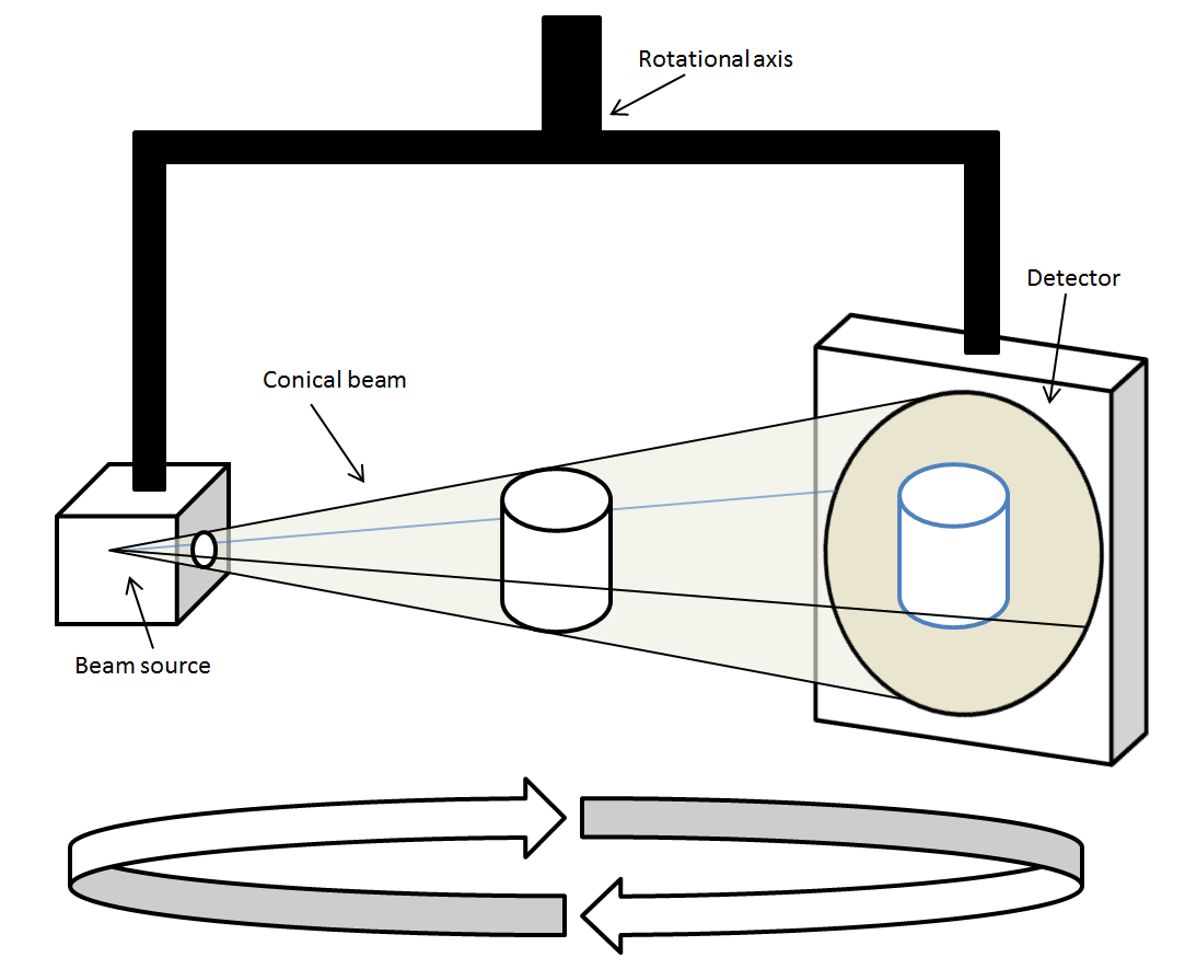
To quickly define 3D dental radiography, also known as Cone Beam or CBCT, it is necessary to understand that unlike 2D scanning, 3D technology makes it possible to have a 3D vision, i.e. to take into account depth. Instead of capturing in pixels, either a square (length x height), radiography makes it possible to capture in voxel, or a cube (length x height x depth). This is made possible by a cone cutting technology rather than a fan cutting technology (see diagram below).

These differences in image capture and the type of result obtained imply a number of advantages and disadvantages that we will define later.
How to x-ray a patient with a cone beam?
Here are a few steps to x-ray a patient using a CBCT:
- Positioning: positioning the patient is much easier with 3D technology, as accuracy has less influence on image quality than with panning.
- Acquisition: Firstly, you must determine the examination area precisely, because the image resolution is much heavier than in 2D, for the same expected image quality, you will probably need a large storage space.
- Result and analysis: the radiography will not give you a single image but a computer file. Ask about the software for reading and processing this data if your X-ray equipment does not come with a workstation. Indeed, export software licenses can be quite expensive.
Advantages and disadvantages of 3D radiography
Advantages of a 3D cone beam
- A single image is sufficient for the entire image of the oral structure
- The practitioner can easily examine the image from several points of view
- Thanks to its pulsed and non-continuous radiation, cone-beam allows the patient to be exposed to X-rays for 10 to 30 times less time
- The cost of an x-ray examination is also a little more profitable for cone-beam (between 80 and 200€) than for scanner (200€ to 380€)
- No storage of films or chemicals
Disadvantages of a 3D cone beam
- Space: an X-ray room is required of at least 3 m2 (for panoramic devices equipped with a 3D function) or 9 m2 (for exclusively 3D devices)
- Price: CBCT has a certain cost but it can be a real investment. It is necessary to count between €100,000 and €200,000 on average, amortized for tax purposes over 7 years
- Technical limitations: it must be understood that for technical reasons, it is not possible to obtain a panoramic view so easily, you will have to build it manually with the software
How to choose a cone beam for your dental practice?
What type of cone beam to choose?
These are the criteria to take into account when choosing your CBCT or Cone-Beam:
Type of image sensor
The image sensor can use the image intensification technique which, thanks to its high sensitivity to light, allows working with low doses of X-rays. However, this type of sensor can be relatively bulky and can generate distortions that must be corrected by the software, thus generating a longer computation time. Flat sensors work just like your phone's camera sensors.
X-ray tube
When it comes to panoramics, the choice of X-ray tube is still very important - you'll have to look at the engine, its characteristics and its power supply
Grayscale
Beyond the possible resolution of the image (counting in pixels for panoramas and voxels for 3D radio), you will have to look at the gray levels through the number of cryptage bits. This setting is important because a wide range of gray levels can differentiate tissues to isolate them.
Image Monitor
Finally, don't forget to see the resolution and quality of your image monitor. Thanks to it you will be able to consult the radiography and deduce the diagnosis, so its quality is paramount.
What do you prioritize according to your dental specialty: orthodontics, implantology, maxillofacial surgery?
The acquisition of a CBCTcan be quite expensive and it is important to choose your equipment according to your needs and, especially, with your specialty. Some devices work better in implant surgery, others are more specialized in pathology diagnosis and oral surgery or endodontics. For example, it is possible to choose a device with a small acquisition field or, conversely, a device with a wider acquisition field..
In orthodontics, the practitioner will probably prefer a wide field of acquisition and opt for a mixed device (panoramic and conical beam). As we have seen before, a virtual radiography reconstituted virtually from a 3D volume, does not offer the same security of interpretation and diagnosis as a panoramic one. But some X-ray machines can automatically switch from the panoramic sensor to the 3D sensor. This option is therefore very interesting in orthodontics.
Beyond the choice of equipment, its use and configuration can vary according to needs. The cut sizes chosen, for example, must be adapted to the type of examination: they will not be the same in orthodontics as in implantology, for example.
To conclude, it is probable that with this article you have understood that the choice of your 3D radiography is important according to your needs and limitations. Finally, while 3D X-rays bring significant benefits to 2D panoramic radio, the latter outperforms 3D technology and remains very practical for certain specific needs.
If you want to learn more about this topic, don't miss this article!Visit our category of Intraoral X-Rays and Panoramic X-Rays and remember to stay informed through our social networks about the latest trends and developments in the dental sector, as well as our discounts, offers, promotions and events. We look forward to seeing you!
Check out the radiology products most recommended by specialists:
All our dental radiology products!