Do you find radiology processes remarkable? We do! From the discovery of X-rays by Wilhelm Conrad Röntgen in 1895 to today's incredible advances such as CBCT technology, it almost feels like no time has passed. In this article we'll tell you all about cone beam computed tomography, also known as CBCT. We'll also explain how it differs from conventional computed tomography (CT) and talk about its very diverse applications in diagnosis and treatment planning in the different areas of dentistry. Let's get started!
Conventional radiology is capable of representing three-dimensional objects in two-dimensional images, but comes with some disadvantages that often hinder accurate interpretation, such as the overlapping and distortion of structures. To overcome these disadvantages, new technologies have emerged, such as conventional computed tomography (CT) and, later on cone beam computed tomography (CBCT).
What is the difference between computed tomography (CT) and cone-beam computed tomography (CBCT)?
CT technology was developed in 1967 by Sir Godfrey Hounsfield and is based on capturing images on the detector screens in multiple planes until a complete image is obtained. This technique exposes the patient to more radiation. The images obtained are useful in maxillofacial trauma, growth and development studies, oral and salivary gland pathology and dental implant planning.
In contrast to CT imaging, in CBCT (Cone Beam Computed Tomography) imaging, the three-dimensional volume of the data is obtained in a single scanner sweep in which the 2D sensor and the radiation source rotate synchronously around the patient's head by 180º or 360º depending on the CBCT equipment used.
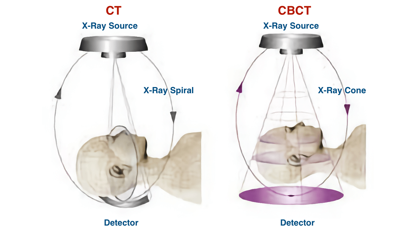
In addition, in a CBCT system, the X-ray beam is a conical shape and produces a cylindrical or spherical data volume, known as the FOV (field of view). The size of the FOV differs between scanners, with some capable of capturing the entire maxillofacial skeleton. Also some CBCT machines can adjust the height of the cylindrical FOV to capture a single area, which reduces the radiation dose to the patient.
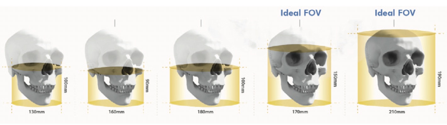
Schematic demonstration of the different sizes of FOV in CBCT.
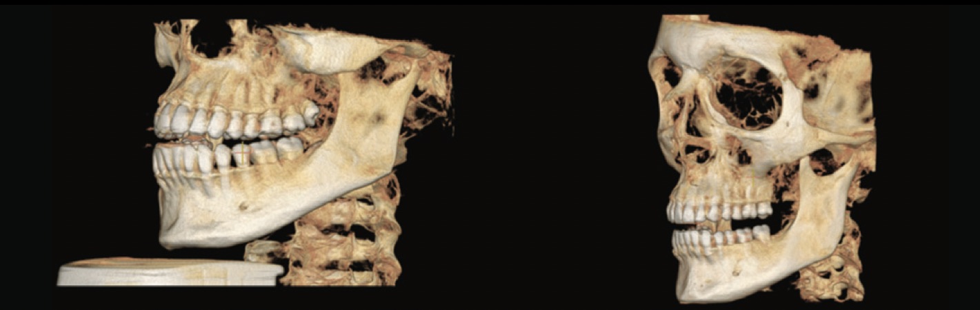
Image comparison: CBCT FOV 16 x 10 versus FOV 15 x 15.
What does CBCT technology provide compared to conventional computed tomography (CT)?
Higher accuracy
Unlike 2D digital images, 3D images are constructed of voxels rather than pixels. The voxel is the smallest element of the 3D radiographic image volume and its size is given by its height, width and depth. In CT the voxels are anisotropic, which means they are not identical in all planes as their height depends on the thickness of the CT beam, i.e. the plane thickness, which means that in certain planes such as the sagittal plane the precision is limited as it depends on the distance of the slices (gap). In contrast, in CBCT the voxels are isotropic, identical in height, width and depth and this allows geometrically more precise images in any plane.
Lower radiation dose
The effective radiation doses of CBCT scanners vary between equipment and configurations in factors such as FOV, but can still be almost as low as in a panoramic X-ray and considerably lower than in a CT scanner. In CBCT the beam is more focused and the radiation is less scattered, so the total radiation would be equivalent to 20% of conventional CT radiation or complete periapical series radiographic exposure.
Applications of CBCT in dentistry: from diagnosis to treatment plan.
According to the FOV we can classify CBCT equipment into large FOV systems (15-30.5 cm) and limited FOV systems (4 to 8 cm). This is important because:
The higher the FOV:
- More extensive imaging of the anatomical area.
- Higher radiation exposure.
- Lower image resolution.
A lower FOV:
- Imaging of a small part of the anatomical area<./li>
- Lower radiation exposure.
- Higher resolution images.
For dentistry applications where maximum detail of structures is not required, but instead a more meaningful representation of the face, such as orthodontics or implant planning, a CBCT scanner with large or moderate FOV may be more effective. But, for example, when diagnosing dental alterations, a more accurate image of a small part is needed, and in this case the use of a limited FOV CBCT system would be more appropriate. Let's take a closer look at the different applications of a CBCT scanner in the dental practice:
Application of CBCT in Implantology
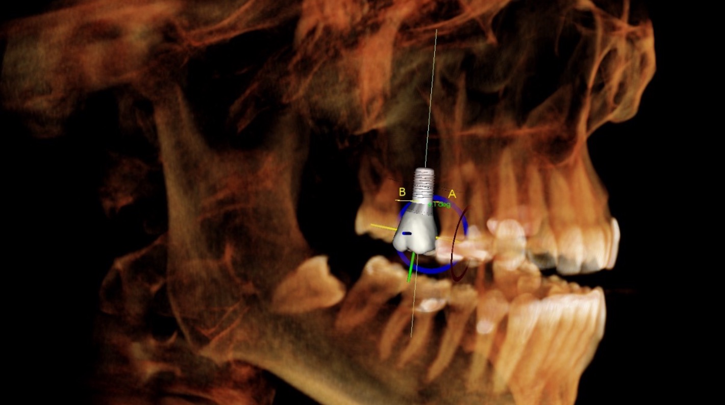
If you use a CBCT scanner to plan your dental implant treatments, some of the benefits would be:
- Determine whether a bone graft or sinus lift is needed.
- Measure the width of the alveolar bone and visualise the contour of the bone.
- Determine the distance between vital anatomical structures.
- Choose the most appropriate implant model and size.
- Optimize implant location and angulation.
- Reduce surgical time.
Application of CBCT in Orthodontics
If you specialise in orthodontics, with a CBCT scanner you'll make the most of great benefits ranging from a more comprehensive orthodontic diagnosis to a more accurate treatment plan. You will be able to perform:
3D cephalometric analysis: forget about the limitations of 2D radiology such as errors in patient positioning, magnification of bilateral structures and the superimposition of other craniofacial structures, and obtain a 3D image of vital structures with better localisation of anatomical marks in cephalometric analysis.
Determination of bone volume, shape and position: if you need to use micro-screws or perform a quick jaw expansion, CBCT will allow you to determine the thickness and shape of the bone in general and in specific areas. In this way you can avoid, for example, placing micro-implants near the roots where they are more likely to fail.
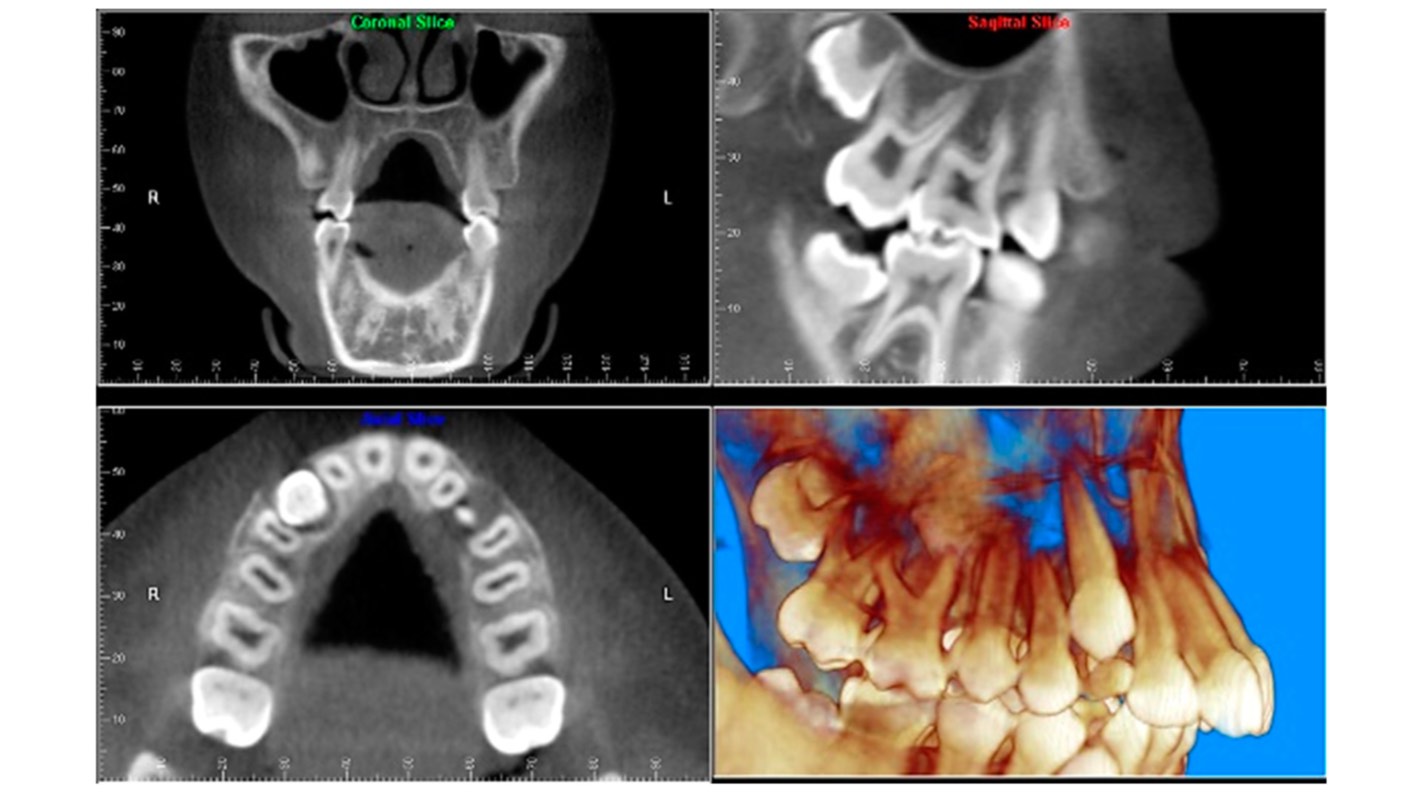
Impactions: CBCT scanners offer greater advantages than conventional radiography when scanning impactions, as they can provide more predictable management and treatment, reducing the risks associated with any impacted tooth. CBCT scanners are also very useful in the accurate diagnosis of the position of supernumerary teeth.
Airway and sinus studies: lateral skull cephalometry is a thing of the past, as with CBCT technology you can improve the volumetric and three-dimensional analysis of the airway.
Morphology of the TMJ: the overlapping of structures such as the petrous region of the temporal bone, the mastoid process and the articular eminence will no longer be a problem. The image quality of the TMJ that you will achieve with your CBCT will be comparable to those obtained with CT, but with the advantage that you can collect this image more quickly and with a lower radiation exposure for the patient. However, magnetic resonance imaging (MRI) remains the gold standard for diagnostic imaging of TMJ.
Application of CBCT in the diagnosis of dental caries
Studies carried out to assess the superiority of CBCT over bitewing, periapical and intraoral radiographs have been performed under well-controlled experimental conditions so do not necessarily reflect the reality of clinical practice given that artefacts in CBCT images of teeth are frequent, mainly in dental crowns. Artefacts are due to metal restorations, implants, endodontic restorative material, among others, and are capable of creating distortion, projecting as light and dark striped lines on adjacent teeth, making correct diagnosis difficult. In conclusion, currently the best radiographic technique for the detection of dental caries is conventional intraoral radiography, despite overestimating its presence.
Application of CBCT in endodontics
If your field of work is endodontics, a CBCT scanner can provide the axial, coronal and sagittal view that you can't achieve with conventional radiology, although conventional radiology is certainly more practical and suitable for the most common endodontic procedures. CBCT has a great advantage in eliminating or reducing the overlapping of structures, for example, for:
Visualising root canal anatomy: if you use a CBCT scanner with a limited FOV you can more accurately recognise root canals and provide more accurate measurements of root angulations.
Identifying periapical pathology: with a CBCT scanner you could obtain greater sensitivity in the diagnosis of experimental periapical lesions, however, we must not forget that artefacts from restorative materials could hinder the diagnosis.
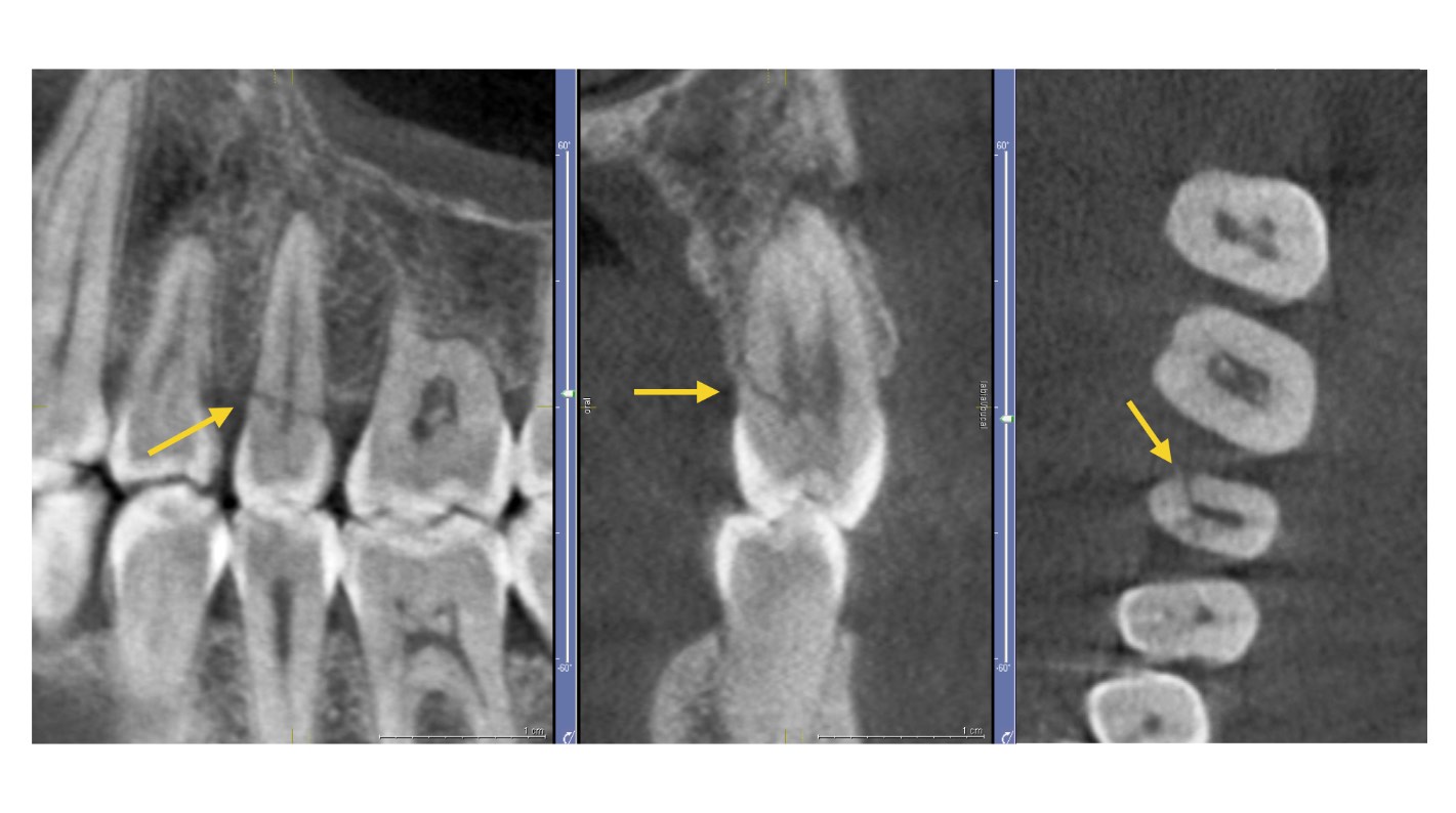
Identification of tooth fractures: the CBCT scanner outperforms conventional radiology in the diagnosis of tooth fractures because unless the beam is oriented so that it passes through the fracture plane, it is not possible to separate the fragments on an intraoral radiograph. Again, this diagnosis on CBCT may be impaired by artefacts.
Analysis of the internal and external root reabsorption process: with your CBCT scanner you can not only detect the exact location, but also determine the extent of the reabsorption and the communication with the periodontal ligament space.
Application of CBCT in periodontics
With a CBCT scanner you will be able to obtain volumetric information of all surfaces. Additionally, you can forget all about the projection limitations of conventional radiology when it comes to determining bone levels in the buccal and lingual areas and also the partial loss of interdental bone thickness.
Application of CBCT in pathology
The CBCT scanner is useful in the detection of cysts, tumours and other anomalies. It is also more sensitive in the detection of invasion due to gingival squamous cell carcinoma, unlike panoramic radiography.
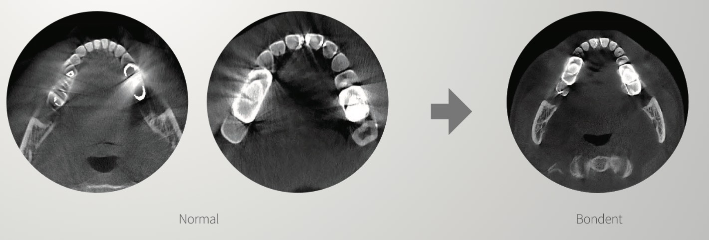
Our recommendation: 1020S CBCT 3-in-1 Dental Scanner (Panoramic, CT and Cephalometric).
One of the most outstanding features of the Bondent CBCT scanner is the intelligent reduction of artefacts, which, as we have seen, make it difficult to obtain a correct diagnosis. In this image you can see the difference:
Other features of the Bondent 3-in-1 CBCT scanner:
- Intelligent noise reduction
- XPI's patented Al algorithm: recognises the maxillary and mandibular dental arches, and also outlines the biological curvature of the teeth automatically.
- Intelligent 3D nano. According to the morphology of the maxilla and mandible, the system automatically generates 33 slices of 0.8 mm thick panoramic CT images with just one touch.
View the Bondent 1020S CBCT 3-in-1 Dental Scanner
In conclusion, a CBCT scanner is a great addition to your dental practice and is very useful for diagnosis and treatment planning in the various specialties of dentistry. It is also a safer option for your patients compared to a CT scanner, as CBCT releases a lower dose of radiation.
No doubt, a CBCT is an expensive piece of equipment, so it's important to read up on all the available information before deciding to buy your own. We hope we have helped you with this blog! However, if you have any questions or concerns, you can contact us through our email and social networks and we'll be happy to help you personally. See you soon!